The Tectorial Membrane
One of the important anatomists of The University of São Paulo, Prof. Olavo Marcondes Calazans, once told me that Anatomy books have many mistakes. This happens because, during his lifetime, an anatomist studies thoroughly only a limited number of subjects. The others he copies from other books. This may help to explain why certain mistakes are sometimes repeated throughout the years.
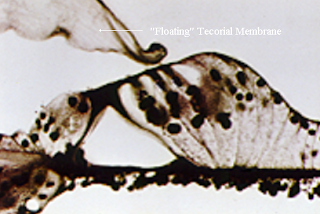
One such example is the tectorial membrane, an important part of the organ of Corti. The section seen in Fig. 1 shows a “floating” tectorial membrane, but this is a fixation artefact.
Anderson Hilding performed precise dissections of the organ of Corti in guinea pigs and stated unequivocally that the tectorial membrane is attached both to the limbus spiralis and to Hensen’s cells. He published this in 1953!
An yet most of the pictures of the inner ear continue to show a floating tectorial membrane (see Fig. 2).
 In 1961 I tried to use different types of preparations in order to get faster fixation. One such substance was acrolein, an unsaturated aldehyde. One characteristic of acrolein fixation was that the tectorial membrane remained attached to the reticular lamina or Hensen’s cells. Some shrinkage of the membrane occurred with fixation but this resulted in bending of the inner pillar rather than in detachment of the tectorial membrane.

A rupture of the tectorial membrane was observed in one of the guinea pigs exposed to noise. The thickening and increased fluorescence of the ruptured margin was proof that it was not an artefact (see Figs. 3 and 4). Rupture of the tectorial membrane seems to be the explanation for profound hearing losses in Menière’s disease, a disorder that does not destroy the hair cells.
Hilding AC. A study of the anatomy and function of the tectorial membrane. Trans Am Acad Ophthalmol Otolaryngol. 1953 Jan-Feb; 57(1): 35-47.
Albernaz PLMA, Covell WP. Acoustic trauma lesions by fluorescence microscopy. Laryngoscope 1962; 72 (10): 1278-96.
One such example is the tectorial membrane, an important part of the organ of Corti. The section seen in Fig. 1 shows a “floating” tectorial membrane, but this is a fixation artefact.

Fig 1. A histological section of the organ of Corti
Anderson Hilding performed precise dissections of the organ of Corti in guinea pigs and stated unequivocally that the tectorial membrane is attached both to the limbus spiralis and to Hensen’s cells. He published this in 1953!
An yet most of the pictures of the inner ear continue to show a floating tectorial membrane (see Fig. 2).

Fig. 2. A drawing of the organ of Corti (Wiki commons)

Fig. 3. A rupture of the tectorial membrane. Fig. 4. Thickening and intense fluorescence at the rupture margin.
A rupture of the tectorial membrane was observed in one of the guinea pigs exposed to noise. The thickening and increased fluorescence of the ruptured margin was proof that it was not an artefact (see Figs. 3 and 4). Rupture of the tectorial membrane seems to be the explanation for profound hearing losses in Menière’s disease, a disorder that does not destroy the hair cells.
Hilding AC. A study of the anatomy and function of the tectorial membrane. Trans Am Acad Ophthalmol Otolaryngol. 1953 Jan-Feb; 57(1): 35-47.
Albernaz PLMA, Covell WP. Acoustic trauma lesions by fluorescence microscopy. Laryngoscope 1962; 72 (10): 1278-96.


Comments
Post a Comment